PARANASAL SINUSES: ANATOMlC TERMlNOLOGY AND NOMENCLATURE
This article is adopted from Annals of Otology, Rhinology and Laryngology Suppl. 167 - Oct 1995 Vol 104, No 10, Part 2, pp7-16
Article Scanned, and Rewritten with a help of OCR system by Kyung Shik Suh, M.D., Dept. of ORL, Ajou University Hospital
EDITED BY
PROF HEINZ R. STAMMBERGER, MD
DAVID W. KENNEDY, MD
Anatomic Illustration coordinated by MAJ WILLLAM E. BOLGER, MC, USAF
FACULTY OF THE ANATOMIC TERMINOLOGY GROUP
PROF HEINZ R. STAMMBERGER, MD, FACILITATOR
MAJ WlLLlAM E. BOLGER, MC, USAF
PROF PETER A. R. CLEMENT, MD
PROF WERNER HOSEMANN, MD
FREDERICK A. KUHN, MD
DONALD C. LANZA, MD
DONALD A. LEOPOLD, MD
TOSHIO OHNISHI, MD
PROF DESIDERIO PASSALI, MD
STEVEN D. SCHAEFER, MD
PROF M. R. WAYOFF, MD
S. JAMES ZINREICH, MD
A consensus on the
preferred modem usage of potentially confusing or ambiguous temls in sinus anatomy and nomenclature is described.
These terms are intended to provide clear communication among otorhinolaryngologists and serve as a basis for discussion among anatomists. Teminology is in English and based on Latin nomenclature. An attempt has been made to reconcile or eliminate duplication, redundancy, and overlap in terminology that have arisen over the past century. A key concept is that the ethmoid complex is divided into anterior and posterior sections by the basal lamella of the middle turbinate.
KEY WORDS-ethmoid sinus, nomenclature, paranasal sinuses, sinusitis, terminology.
lNTRODUCTION
Recent advances in clinical technology, especially computed tomography (CT) and microscopic and endoscopic sinonasal surgery, have given renewed importance to the standardization of anatomic terminology in sinusitis. Current understanding of the localization and extent of the pathophysiology of sinus and skull base disease is based on detailed knowledge of anatomic structure. Because the opportunities for intervention are virtually unprecedented in their precision, it is imperative that surgeons and radiologists cornmunicate as efficiently and as accurately as possible.
Much of the confusion and variation in nomenclature and terminology, as well as some central issues in the pathophysiology of sinus disease, surround the structures related to the ethmoid sinuses.l The terminology in this article reflects preferred modern usage of important anatomic terms as recommended in the reports of the Anatomic Terminology Group, developed at the Intemational Conference on Sinus Disease: Terminology, Staging, and Therapy, held in the United States in Princeton, New Jersey, in July 1993. Kaufmann's4 "double middle turbinate"(Gedoppelte mittlere Muschel) describes the medially bent uncinate process that curves out of the middle meatus like the brim of a hat. The structure is not, however, a double middle turbinate in reality. Kaufmann' s term may be retained for its historical interest but not used in a rigorous anatomic context. The same applies to variations such as a paradoxicallycurved middle turbinate.
- The nomenclature and anatomic concepts presented for the lateral nasal wall and anterior skull base evolved over several decades and have proved to be satisfactory for endoscopic and microscopic diagnosis and surgery as well as for radiology.
- Topographic and directional instructions(such assuperior and inferior) are given relative to a standing person.
BEHIND THE CONFUSION IN ETHMOID TERMINOLOGY
Confusion and disparate practices exist today concerning the definitions and usage of such terms as hiatus semilunaris, infundibulum, frontal recess, and nasofrontal duct. It is interesting to note, however, that many of the misunderstandings date from the time when the terms were first coined. While Zuckerkandl5,6 used the term hiatus semilunaris in much the same way as we do, to signify the two-dimensional cleft between the posterior margin of the uncinate process and the anterior face of the ethmoid bulla, the term ethmoid infundibulumhas been used more loosely.
Anatomists today consider the ethmoid infundibulum to be a three-dimensional structure that is actually a well-defined cleft. Initially, however, Zuckerkandl applied the term only to the depression extending forward from the hiatus semilunaris both inferiorly and superiorly into the lateral nasal wall. He attributed the term to Boyer, even though what Boyer described was the cleft that Killian7 later called the frontal recess. Since then, others have erroneously used the terms frontal infundibulum and nasofrontal duct synonymously with frontal recess.
Additional terms for the clefts of the anterior ethmoid region have been invented and applied without regard to consistency. As a result, numerous terms have come into being for a single structure. For example, what was termed recessus frontalis by one author was called ductus nasofrontalis and recessus anterior meatus medii by later contributors to the literature. For another example, the frontal recess has been called the frontal infundibulum of the hiatus semilunaris, and the terms hiatus semilunaris and ethmoid infundibulum have been used for the same structure.
ETHMOID COMPLEXES
The structures of the lateral nasal wall and paranasal sinuses fall into two anatomically and physiologically distinct categories, the anterior and posterior ethmoidcomplexes (Fig 1) PNS1.gif. . The basal lamella of the middle turbinate is the clear and distinct separation between thetwo ethmoid complexes, according to definition, patterns of mucociliary secretion transport, and embryologic development. Cells and clefts that open and drain anteriorly and inferiorly to this lamella belong to the anterior ethmoid complex; those that open or drain posteriorly or superiorly, with the exception of the sphenoid sinus, belong to the posterior ethmoid complex. The expressions middle ethmoid and middle ethmoid cells should not be used, because in terms of anatomy, physiology, or function, no structure represents the middle of the ethmoid complex.
BASAL LAMELLA OF MIDDLE TURBINATE
This structure is actually the third basal lamella of the ethmoturbinals (Fig 2) PNS2.gif . The most anterior and superior insertion of the middle turbinate is adjacent to the crista ethmoidalis of the maxilla. The posterior end is attached to the crista ethmoidalis of the perpendicular process of the palatine bone(lamina perpendicularis).
The area between comprises three parts . The anterior third of the middle turbinate inserts vertically into the skull base at the lateral edge of the lamina cribrosa. The middle third turns laterally across the skull base to the lamina papyracea, where it turns inferiorly. The most posterior segment becomes horizontal.
The insertion of the middle turbinate thus lies in three different planes. The anterior segment lies sagittally, attaching to the lateral end of the lamina cribrosa opposite its lamina lateralis. The middle segment is fixed to the lamina papyracea in an almost frontal plane. The posterior segment is attached to the lamina papyracea, the medial wall of the maxillary sinus, or both, to form the roof of the posterior third of the middle meatus.
The stability of the middle turbinate accrues largely from its fixation along three planes. The frontal and posterior portions, which are vertical and horizontal, respectively, are part of the basal lamella of the middle turbinate. The middle section, also part of the basal lamella, is not necessarily a smooth surface. Cells or well-pneumatized clefts of the anterior ethmoid can indent this plate dorsally, giving it a posterosuperior orientation. When a retrobullar recess is well developed, anterior ethmoid cells can reach back almost to the sphenoid sinus. Conversely, cells of the posterior ethmoid or the superior meatus can create an anterior bulge in the midsection of the basal lamella.
[Discussion. The turbinates originate from the lateral nasal wall during development, and each has its own basal lamella. As the basal lamella of the third ethmoturbinal, which separates the anterior and posterior ethmoid complexes, the middle turbinate is anatomically and physiologically the most important ofthe basal larnellas. Because there is more than one basal lamella, however, when the term basal lamella is used, the turbinate with which it is associated must bespecified.
The role of the basal lamella of the middle turbinate as the division between anterior and posterior ethmoid complexes is underscored by the French terminology, racine cloisonnante du cornet moyen, which means the dividing root of the middle turbinate. The term basal is preferable to ground to avoid misnomers such as grand lamella, which lend themselves to misconceptions and confusion.]
ANTERIOR ETHMOID AND RELATED STRUCI'URES
Uncinate Process.
The term derives from the Latin, processus uncinatus, meaning hooked outgrowth, and refers to a remnant of the descending portion of the first ethmoturbinal.
The uncinate process is a thin, bony leaflet that resembles a hook (Fig 3) PNS3.gif . It is oriented almost sagittallyand runs from anterosuperior to posteroinferior. Its concave posterosuperior free margin is parallel to the anterior surface of the ethmoid bulla. The uncinate process attaches to the perpendicular process(lamina perpendicularis) of the palatine bone and the ethmoid process of the inferior turbinate with bony spicules. The convex anterior margin ascends to the lacrimal bone,and sometimes to the skull base or lamina papyracea, remaining in contact with the bony lateral nasal wall(Fig 4) PNS4.gif . When curved medially to a greater than usual extent, the free margin of the uncinate process may protrude into, and sometimes even out of, the middle nasal meatus. The uncinate process may attach to the middle turbinate superiorly, too, when curved medially in its superior most portion. In rare cases, the superior part of the uncinate process may attach with several "fingers" to the middle turbinate, the skull base, and the lateral nasal wall as well.
Agger Nasi.
The term comes from the Latin for nasal mound and refers to the most superior remnant of the first ethmoturbinal, which persists as a mound or crest immediately anterior and superior to the insertion of the middle turbinate (Fig 5) PNS5.gif . An agger nasi cell results when this area of the lateral nasal wall under goes pneumatization. Depending on the degree of pneumatization, agger nasi cells may reach laterally to the lacrimal fossa and cause narrowing of the frontal recess.
Ethmoid Bulla.
From the Latin, bulla ethmoidalis,where bulla means a hollow, thin-walled, bony prominence, the name refers to the largest and most nonvariant air cells in the anterior ethmoid complex. It is formed by pneumatization of the bulla lamella, or second ethmoid basal lamella, and is like a bleb on the lamina papyracea. The bulla lamella can form the posterior wall of the frontal recess if it reaches the roof of the ethmoid. Failure to reach the skull base, however, results information of the suprabullar recess, an aerated space of varying dimensions between the bulla lamella and the skull base.
[Discussion. The ethmoid bulla, created when the second basal lamella of the ethmoturbinals is pneumatized, is sometimes called a promontory in the literature. In the absence of pneumatization, it does not exist. The traditional anatomic term for a persisting and nonpneumatized second basal lamella is torus ethmoidalis. The suggestion to change the name torus bullaris was rejected by the Anatomic Terminology Group because the term is oxymoronic. Torus describes a solid structure, and bullaris refers to apneumatized structure. The term torus lateralis does not specifically denote the non-pneumatized bulla lamella.]
Suprabullar and Retrobullar Recess(Sinus Lateralis).
The Latin term recessus suprabullaris et retrobullaris has as synonyms the sinus lateralis of Grunwald8 and the susbullar cell of Mouret.9,l0 The suprabullar recess may extend into a retrobullar recess if the posterior wall of the bulla lamella is not in contact with the basal lamella of the middle turbinate. When well developed, this space is bordered superiorly by the ethmoid roof, laterally by the lamina papyracea, inferiorly by the roofof the ethmoid bulla, and posteriorly by the basal lamella of the middle turbinate. Anteriorly, it is separated from the frontal recess only when the bulla lamella reaches the skull base . Otherwise, the suprabullar recess opens into the frontal recess. The suprabullar and retrobullar recess also can be approached medially and inferiorly through the hiatus semilunaris superior.
[Discussion . This space does not have a single openingfor ventilation and drainage, and therefore does notsatisfy the criteria of a cell. The term recess is recommended because the space can be approached anteriorly and superiorly from the frontal recess and medially and inferiorly from the hiatus semilunaris superior. Grunwald's term, sinus lateralis, is suitable anatomically, but the complete term, sinus lateralis sinus ethmoidalis, is necessary to differentiate it from the lateral sinus in the brain. The latter term is considered too long to be practical.]
Hiatus Semilunaris Inferior.
The origin of this termis the hiatus semilunaris inferior of Grunwald.8 The hiatus semilunaris inferior is an anatomic plane that represents the shortest distance between the free posterior margin of the uncinate process and the corresponding anterior face of the ethmoid bulla. Typically, but not necessarily, it lies in the sagittal plane and does not represent a true space (Fig 3) PNS3.gif .
Two concepts are helpful when considering the term hiatus semilunaris. First, the Latin root translates directly into English as cleft, gap, or passageway; indeed, the hiatus semilunaris inferior is a crescent-shaped cleft. Second, the passageway is like a doorway through which one must pass to arrive at the ethmoid infundibulum, which is a three-dimensional space. Hiatus Semilunaris Superior. This is a second, but only vaguely defined, crescent-shaped cleft between the ethmoid bulla and the middle turbinate. The suprabullar and retrobullar recess can be entered medially and inferiorly underneath the middle turbinate through the hiatus semilunaris superior.
[Discussion. The term inferior rather than anterior is recornmended for the hiatus semilunaris that is located between the uncinate process and the ethmoid bulla,and superior rather than posterior is recommended for the hiatus semilunaris that may lead into the suprabullar and retrobullar recess. When the orientation of the head conforms to that of a standing person, inferior and superior are more accurate.]
Infundibulum.
The term infundibulum (plural, infundibula) connotes a funnel shaped structure and comes from the Latin infundere, meaning to pour into. There are three different infundibula in the paranasal sinuses: the frontal, maxillary, and ethmoid. The ethmoid infundibulum is the most important pathophysiologically, and the others are notable primarily for historical perspective. The ethmoid infundibulurn, from infundibulum ethmoidale, is a cleft or true three-dimensional space. Were a cast made of the space, it would typically resemble an inverted segment of grapefruit with the wide edge facing posteriorly.
The ethmoid infundibulum is bordered medially by the uncinate process and laterally by the lamina papyracea. The frontal process of the maxilla and the lacrimal bone may constitute parts of the lateral wall anterosuperiorly, but this is rare. Fusion with the anterior border of the uncinate process provides a connection with the inferior turbinate.
At its anterior end, the ethmoid infundibulum ends blindly in an acute angle, giving rise to the V-like shape noted in axial sections and on CT scans. Posteriorly, the ethmoid infundibulum extends to the anterior face of the ethmoid bulla and opens into the middle meatus through the hiatus semilunaris inferior. Periosteum and mucous membrane cover bony defects in the lateral nasal wall, forming the anterior and posterior nasal fontanelles.
The maxillary sinus ostium usually can be found at the floor and lateral aspect of the infundibulum between its middle and posterior third. From the middle meatus, the natural ostium of the maxillary sinus therefore remains hidden, lateral to the uncinate process in the ethmoid infundibulum.
The relationship between the ethmoid infimdibulum and the skull base, especially the frontal recess, depends on the uncinate process. Superiorly, the ethmoid infundibulum may end blindly in the terminal recess, or recessus terminalis, if the uncinate process bends laterally and inserts onto the lamina papyracea. If the uncinate process reaches to the skull base or fuses with the middle turbinate medially, the ethmoid infundibulum may pass into the frontal recess superiorly.
[Discussion. The frontal sinus and maxillary sinus infundibula are inside their respective sinus cavities and resemble narrowing or funneling tunnels toward their natural ostia. The border between the ethmoid infundibulum and the frontal recess is difficult to define. Embryologically, the ethmoid infundibulum and frontal recess arose from a single structure, and the variations of the uncinate process determine the relationship between the two in maturity.]
The frontal infundibulum, named from the Latin infundibulum sinus frontalis or infundibulum frontale, is a funnel-shaped narrowing of the inferior aspect ofthe frontal sinus toward the floor of the frontal sinus ostium. It is located inside the frontal sinus. The maxillary infundibulum, after the Latin infundibulum sinus maxillaris or infundibulum maxillare, is the funnel-shaped narrowing of the lumen of the maxillary sinus toward its natural ostium. Typically, the lumen does not narrow significantly toward the maxillary sinus ostium.
Frontal Recess.
Perhaps the most complicated structure in the anterior ethmoid complex, the frontal recessis the most anterior and superior portion of the complex that leads to and communicates with the frontal sinus (Fig 5) PNS5.gif . It is not synonymous with the nasofrontal duct.
The medial wall of the frontal recess is the most anterior and superior part of the middle turbinate. The lateral wall is mostly lamina papyracea. A discrete posterior margin exists only when the basal lamella ofthe bulla reaches the skull base, separating the frontal recess from the suprabullar recess . If the insertion of the bulla lamella reaches far anteriorly and/or the bulla is well pneumatized, the frontal recess becomes narrowed from the posterior. This may result in a tubular appearance on sagittal section, which is the reason the narrowed recess came to be known, albeit incorrectly, as the nasofrontal duct. Under certain conditions, a tubular structure can be the communication between the frontal recess and the frontal sinus.
In sagittal section, the frontal recess usually has the shape of an inverted funnel. When taken together withthe frontal infundibulum, the shape resembles an hourglass, with the constricted portion being at the level of the natural ostium of the frontal sinus. The floor of the frontal recess varies so much that it has no uniform definition.
[Discussion. Although the anatomic definitions are clear, there is great confusion in usage among these terms. The frontal recess is the most anterior and superior part of the anterior ethmoid complex. From here, the frontal bone becomes pneumatized, resulting in a frontal sinus. Seen from above, the frontal recess narrows toward its ostium (through the frontal infundibulum). From the level of the ostium, the frontal recess then widens in the inferior and posterior direction, usually in the shape of an inverted funnel.
When this communication is narrowed from behind by the ethmoid bulla or the bulla lamella or from in front by a pneumatized agger nasi cell,a short, ductlike structure results. The bony walls of the resulting structure are not truly its own, however, so to call it a duct or other tubular structure is not anatomically correct. Its ductlike appearance on sagittal or coronal CT scans is misleading.
The formation of additional cells in the frontal recess and the infundibulum, apart from agger nasi cells, is highly individual. The Anatomic Terminology Group recommends that they be described according to their anatomic orientation. For example, if they reach the lacrimal sac and pneumatize into the lacrimal bone, they would be lacrimal cells of the ethmoid infundibulum or lacrimal cells of the frontal recess. A cell that pneumatizes into the frontal bone is likewise a frontal cell of the anterior ethmoid or a bulla frontalis. Terms such as threshold cell are to be avoided. A supraorbital cell is an anatomic variant that develops as an extension, from the posterior aspect, of the frontal or suprabullar recess. It is therefore called a supraorbital cell of the frontal recess or a supraorbital cell of the suprabullar recess.]
Nasal Fontanelles.
These are the areas of the lateral nasal wall in which no bone exists. They are usually found immediately above the insertion of the inferior turbinate. Thus, the mucosa of the maxillary sinus and the middle meatus are separated only by a fibrous layer of periosteum. The fontanelles may be sites of accessory ostia to the maxillary sinus. The anterior fontanelleis inferior and anterior to the uncinate process; the posterior fontanelle is superior and posterior to the part of the uncinate process that fuses with the medial wall of the maxillary sinus.
Roof of Ethmoid.
Lateral to the lamina cribrosa and to the inserlion of the middle turbinate, the ethmoid bone is open superiorly. The ethmoid roof itself is created by the frontal bone. Indentations or foveolae in the frontal bone cover the corresponding clefts and cells ofthe ethmoid.
Keros11 has described three different and surgically important types of configurations of the ethmoid roof(Fig 6) PNS6.gif . The differentiation depends on the length of thelateral lamella of the cribriform plate, which is the thinnest bone in the entire anterior skull base.l2 In type 1, the olfactory fossa is only 1 to 3 mm deep, the lateral lamella is short (almost nonexistent), and the ethmoid roof is almost in the same plane as the cribriform plate. In type 2, the olfactory fossa is from 4 to 7 mm deep, andthe lateral lamella is longer. In type 3, the olfactory fossais 8 to 16 mm deep, and the ethmoid roof lies significantly above the cribriform plate. Because of the danger that instrumentation can penetrate the thin and vulnerable lateral lamella, this is the configuration of greatest concern for the surgeon.
[Discussion. Controversy surrounds the terms fovea and foveolae ethmoidales. The anterior two thirds of the ethmoid complex opens superiorly, and the cells and clefts in this area are closed over by the frontal bone. Small pits or indentations overlie the open ethmoid clefts and spaces, which are open superiorly. These indentations are the foveolae (from the Latin, foveolae ethmoidales ossis frontalis, meaning ethmoid pits of the frontal bone). Use of the term fovea for the entire ethmoid roof doesnot distinguish between the endonasal view and the view from above looking down on the olfactory groove. While any individual pit may be called a fovea or foveola, the ethmoid roof may not. The Anatomic Terminology Group recommends that only the term foveolae ethmoidales(of the frontal bone) be used and advises that the terms fovea and dome of the ethmoid not be used. The clinical significance of the Keros classification accrues from the fact that the risk of intracranial entry during surgery increases with the length and, consequently, angulation of the lateral lamella of the cribriform plate. When a patient has a type 3 configuration,perhaps 14to 16mm of anterior cranial fossa is medial to a place where instrumentation may be used.]
Concha Bullosa.
When there is pneumatization of the middle turbinate, the term concha bullosa is used. The term may also apply to pneumatization of the superior turbinate. The pneumatization of the middle turbinate usually originates from the frontal recess or the agger nasi, and growth of a concha bullosa may begin late in life.
[Discussion. The concha bullosa must be distinguished from an interlamellar cell (Fig 7) PNS7.gif , which arises from pneumatization of the vertical lamella of the middle turbinate from the superior meatus . The concha bullosa is a normal variant that in itself does not require surgery, but the presence of a concha bullosa may predispose a patient to occlusion of the ostiomeatal complex and subsequent sinus disease. Therefore, surgery may be appropriate.]
Infraorbital Ethmoid Cell (Haller's Cell).
The potential pathophysiologic importance of a Haller' s cell is clear, but the anatomic definition is not. As described by Haller in 1765, these cells grow into the bony orbital floor that constitutes the roof of the maxillary sinus, are differentiable from the bulla, and have a potential pathophysiologic relationship to a narrowed ethmoid infundibulum or maxillary sinus ostium (Fig 8) PNS8.gif .
The term cellula orbitoethmoidalis or orbitoethmoidcell does not indicate, for example, that the cell grows directly into the floor of the orbit. The term infraorbitalethmoid cell is better because it implies contrast with a supraorbital cell that originates from the frontal or suprabullar recess. For exactitude, the full term is infraorbital cell of the anterior or posterior ethmoids, depending on its origin.
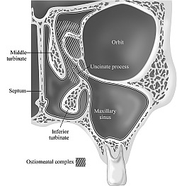
Ostiomeatal Complex.
No consensus exists to define exact anatomic descriptions of the borders and margins of the ostiomeatal complex.l3 Rather, the ostiomeatal complex is a functional entity of the anterior ethmoid complex that represents the final common pathway for drainage and ventilation of the frontal, maxillary, and anterior ethmoid cells (Fig 9) PNS9.gif . Any or all cells, clefts, and ostia, with their dependent sinuses, may become diseased, thereby contributing to the symptoms and pathophysiology of sinusitis.
POSTERIOR ETHMOID AND RELATED STRUCTURES
Posterior Ethmoid and Sphenoid Sinus .
There are only a few terms in anatomic nomenclature that require a better definition and explanation in these regions.
Like the middle turbinate, the superior and, if present, supreme turbinates are attached to the lateral nasal wall and the anterior skull base by means of their basal lamellas. The course of attachment of the supreme turbinate is similar to that of the middle turbinate but of less significance in pathophysiology and surgery. The superior nasal meatus (meatus nasi superior) and supreme nasal meatus(meatus nasi supremus) lie underneath the respective turbinates. Because the individual cells and clefts underneath the supreme(or fourth) turbinate are nowhere defined in the literature, they are considered part of a single posterior ethmoid complex.
The sphenoethmoid recess (recessus sphenoethmoidalis) is the space between the superior (and supreme, if present) turbinate laterally, the roof of the nose(rima olfactoria) superiorly, and the nasal septum medially. Its posterior border is the anterior face of the sphenoid bone. Medially there is no clearcut inferior border, and laterally the inferior border is seen at the inferior margin of the superior turbinate. The anterior extension is equally ill-defined, passing into the common nasal meatus.
Sphenoethmoid Cell.
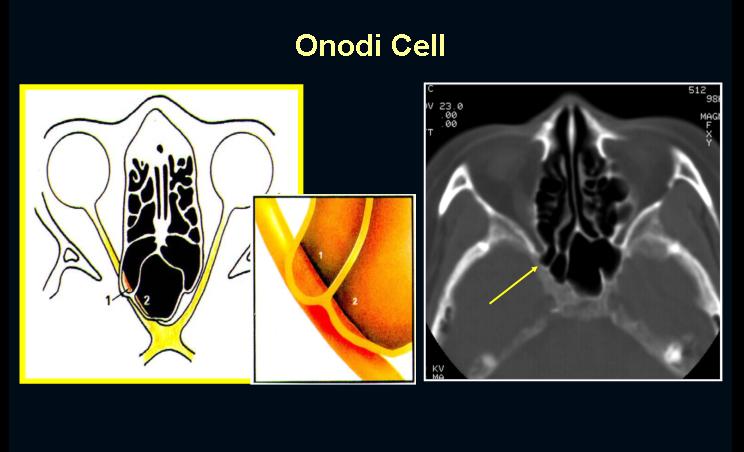
Posterior ethmoid cells can become pneumatized far laterally and to some degree superiorly to the sphenoid sinus, in which case they are called sphenoethmoid cells(cellulae sphenoethmoidales) or Onodi cells (Fig 10) PNS10.gif . Pneumatization of the clinoid process in those cases may originate from the posterior ethmoid cell, also.
The optic nerve and carotid artery may be exposed in a sphenoethmoid(Onodi) cell. This is clinically significant because the sphenoid sinus is located medially and inferiorly to the most posterior cell of the posteriorethmoid complex. Consequently, attempts to use instrumentation to locate the sphenoid sinus directly behind the last cell of the posterior ethmoid complex may result in serious damage to the optic nerve or carotid artery.
[Discussion. The most posterior ethmoid cell may becalled a sphenoethmoid cell(Onodi cell) when it pneumatizes laterally and superiorly to the sphenoid sinus and is intimately associated with the optic nerve. Prominence of the optic nerve tubercle or the internal carotid artery is not prerequisite, however. Moreover, the optic nerve tubercle may be prominent in other posterior ethmoid cells as well. Whether ethmoid complex components grow posteriorly alongside the sphenoid sinus or sphenoethmoid cells pneumatize directly into the sphenoid bone has not been resolved, but the answer does not bear on practical issues in diagnosis and sugery: the air space in question isclearly ethmoid.]
Optic Nerve Tubercle.
The bulge of the medial aspect of the bone surrounding the optic foramen (foramen opticum) is the optic nerve tubercle (tuberculum nervi optici) . Depending on the degree of pneumatization and the presence and configuration of sphenoethmoid cells, it can be seen in the posterior ethmoid cells, at the transition between the posterior ethmoid and sphenoid sinuses, or in the sphenoid sinus itself. The optic canal (canalis opticus) can project into the sinus lumen, and dehiscences of the bony wall may be present. The optic nerve may pass through a sphenoethmoid cell or sphenoid sinus like a column, surrounded by pneumatized space. There may be an infraoptic recess between the opticnerve and the intemal carotid artery. The more pronounced the pneumatization of the anterior clinoid process, the deeper the recess.
EMBRYOLOGY OF BONY LATERAL NASAL WALL
Understanding the embryologic development of the turbinates and the ethmoid sheds light on the complex relationships involving the basal lamella. The ethmoid turbinates form on the lateral nasal wall of the fetus, giving rise to six ridges during the 9th and 10th weeks of fetal development. These typically fuse into fewer ridges, which become clearly separated by furrows. Like mature turbinates, each ridge and furrow has an anterior ascending and a posterior descending portion.
During development, the first ethmoturbinal regresses and does not become permanent. The descending portion of the first ethmoturbinal remains as the uncinate process,and the ascending portion remains asthe agger nasi (ie, the nasoturbinal). The middle and posterior sections of the depression between the first and second ethmoturbinals(the descending portion) become the ethmoid infundibulum, and the superior ascending part becomes the frontal recess. The frontal sinus is formed by pneumatization of the frontal recess into the frontal bone. The inferior turbinate, known as the maxilloturbinal, is a single bone unrelated to the ethmoturbinals.
From these embryologic relationships, one can see that the uncinate process is actually the basal lamella of the first ethmoturbinal. Similarly, the ethmoid bulla evolves from the second basal lamella, and the middle turbinate from the third basal lamella.
Given the variety of anatomic features of an ethmoid cell, the only fixed point of reference is the ostium. Thus, cells are classified as belonging to the anterior ethmoid when they drain into the middle meatus and belonging to the posterior ethmoid when they drain into the superior meatus.
The phrases ethmoid cells of the middle meatus and ethmoid cells of the superior meatus may be more strictly correct than the terms anterior ethmoid sinus and posterior ethmoid sinus, which are currently in use. Such a major change in nomenclature, however, could be confusing. Therefore, we continue to use the terms anterior ethmoid and posterior ethmoid and designate the basal lamella of the middle turbinate as their separation.
CONCLUSION
The Anatomic Temlinology Group's suggestions for a unified system of terminology are designed to provide a current intenational language for otorhinolaryngologic surgeons and to serve as a basis for discussion among anatomists. The Group plans to renew its discussions of anatomic terminology and nomenclaturein 1997.
REFERENCES
INTRODUCTION
The complexity of the paranasal sinuses anatomy, as well as their many functions make the sinuses an interesting and rewarding topic of study. There are a total of four paired sinuses. They include the frontal, ethmoid, maxillary and sphenoid sinuses. These sinuses are essentially mucosa-lined airspaces within the bones of the face and skull. Their development begins in the womb, but results in only two clinically-relevant sinuses by birth--the maxillary and ethmoid sinuses. The development of the lateral nasal wall begins with a smooth, undifferentiated structure. The first outgrowth is the maxilloturbinal which will eventually become the inferior turbinate. Subsequently, another mound of mesenchyme forms which is the ethmoturbinal, destined to become the middle, superior, and supreme turbinates by subdividing into the second and third ethmoturbinals. This growth is followed by the development of the agger nasi cells, uncinate process, and ethmoid infundibulum. The sinuses then begin to develop. The resultant system of cavities, depressions, ostia, and processes is a complex system of structures which must be understood in detail before surgical management of sinus disease can be safe and effective. The physical anatomy, microscopic anatomy, physiology and function of the sinuses will be explored.
LATERAL NASAL WALL
The lateral nasal wall includes portions of the ethmoid, the maxilla, the palatine, the lacrimal, the medial pterygoid plate of the sphenoid, the nasal and the inferior turbinate bones. Three to four turbinates project from the wall; the supreme, superior, and middle turbinates being projections of the ethmoid bone. The inferior is considered to be an independent structure. Each of these structures overlies an airspace beneath and lateral to it known as a meatus. A small slip of bone projects from the ethmoid bone which covers the opening(s) of the laterally placed maxillary sinus and forms a trough behind the middle turbinate. This thin bony partition is known as the uncinate process. The superior nasal sidewall consists of ethmoid sinus cells which laterally border the olfactory epithelium and the fragile cribiform plate. Superior to the anterior most ethmoid cells lies the frontal sinus which drains between an assortment of air cells. The posterior-superior aspect of the lateral nasal wall becomes the anterior wall of the sphenoid sinus which nestles below the sella turcica and the cavernous sinus.
MAXILLARY SINUS
Development
The maxillary sinus (antrum of Highmore) is the first to develop. These structures are usually fluid-filled at birth. The growth of these sinuses is biphasic with growth during years 0-3 and 7-12. During the later phase pneumatization spreads more inferiorly as the permanent teeth take their place. Pneumatization can be so extensive as to expose tooth roots with only a thin layer of soft tissue covering them.
Structure
The adult maxillary sinus is a pyramid which has a volume of approximately 15 ml (34x33x23mm). The base of the pyramid is the nasal wall with the peak pointing toward the zygomatic process. The anterior wall has the infraorbital foramen located at the midsuperior portion with the infraorbital nerve running over the roof of the sinus and exiting through the foramen. This nerve can be dehiscent (14%). The thinnest portion of the anterior wall is just above the canine tooth--the canine fossa. The roof is formed by the orbital floor and transected by the course of the infraorbital nerve. The posterior wall is unremarkable. Behind this wall is the pterygomaxillary fossa with the internal maxillary artery, sphenopalatine ganglion and the Vidian canal, the greater palatine nerve and the foramen rotundum. The floor, as discussed above, varies in it's level. From birth to age nine the floor of the sinus is above that of the nasal cavity. At age nine the floor is generally at the level of the nasal floor. The floor continues to sink as the maxillary sinus pneumatizes. Because of the close relationship with the dentition dental disease can cause maxillary infection, and tooth extraction can result in oral-antral fistulae.
Vascular supply
Branches of the internal maxillary artery supply this sinus. These include the infraorbital (as it runs with the infraorbital nerve), lateral branches of the sphenopalatine, greater palatine, and the alveolar arteries. Venous drainage runs anteriorly into the facial vein and posteriorly into the maxillary vein and jugular vs. dural sinus systems.
Innervation
The maxillary sinus is innervated by branches of V2. Specifically, the greater palatine nerve and the branches of the infraorbital nerve.
Related structures
Nasolacrimal duct
The nasolacrimal duct drains the lacrimal sac and runs from the lacrimal fossa in the orbit down the posterior aspect of the maxillary vertical buttress and empties in the anterior aspect of the inferior meatus. The duct lies very close to the maxillary ostium. On average it lies 4mm-9mm anterior to the ostium.
Natural ostium
The natural maxillary ostium is located at the superior aspect of the medial wall of the sinus. Intranasally, it is usually in the posterior half of the ethmoid infundibulum, or behind the lower 1/3 of the uncinate process. The posterior edge of the ostia is continuous with the lamina papyracea, thus a reliable landmark for the lateral limit of surgical dissection. The ostium size averages 2.4 mm but can vary from 1 to 17mm. The ostium is much smaller than that actual bony defect, as mucosa fills this area and defines the extent of the opening. 88% of maxillary ostium are hidden behind the uncinate process and therefore cannot be visualized endoscopically.
Anterior/Posterior Fontanelles/Accessory Ostium
Two bony dehiscences of the lateral nasal wall/maxillary sinus medial wall exist (sometimes there is one large bone dehiscence. These are usually covered by mucosa. In some individuals the anterior or posterior fontanelles may be patent which results in an accessory ostium. They are nonfunctional ostia and serve to drain the sinus only if the natural ostium is blocked and intrasinus pressure/gravity moves material out of the ostium. Accessory ostium are usually found in the posterior fontanel.
ETHMOID SINUSES
Development
The ethmoid sinuses are well-delineated, fluid-filled structures in a newborn child. During fetal development the anterior cells form first, followed by the posterior cells. The cells grow gradually and are adult size by age 12. They are not usually seen on radiographs until age one. The septa gradually thin and pneumatization spreads as the child ages. Ethmoid cells are the most variable and can often be found above the orbit, lateral to the sphenoid, into the roof of the maxillary sinus, and anteriorly above the frontal sinus. These cells have been named. A cell above the orbit is called a supraorbital cell and is found in 15% of patients. Invasion of an ethmoid cell into the floor of the frontal sinus is called a frontal bulla. Extension into the middle turbinate is termed concha bullosa. Cells in the roof of the maxillary sinus (infraorbital) are called, "Haller's cells," and are found in 10% of the population. These cells can obstruct the maxillary ostia and narrow the infundibulum and result in disruption of normal sinus function. Finally, a cell which extends anteriolaterally to the sphenoid sinus is called an Onodi cell (10%). The common variability of these cells makes preoperative imaging essential to clarify a patient's individual anatomy.
Structure
Posterior and anterior cells combined have a volume of 15 ml (3.3x2.7x1.4cm). The ethmoids are shaped like a pyramid and are divided into multiple cells by thin septa. The roof of the ethmoids is composed of multiple important structures. The roof slopes both posteriorly (angle of 15 degrees) and medially. The anterior 2/3 of the roof is thick and strong and is composed of the frontal bone and the foveolae ethmoidalis. The posterior 1/3 is higher laterally and slopes down medially to the cribiform plate. The junction between the lateral dense bone and the plate is one-tenth as strong as the lateral roof. The difference in height between the lateral and medial roof is variable, but can be as much as 15-17mm. The posterior aspect of the ethmoid cells borders on the sphenoid sinus. The lateral wall is the lamina papyracea of the orbit.
Vascular supply
The ethmoid sinuses are supplied by blood flow originating from both the external and internal carotid arteries. The Sphenopalatine artery as well as the ophthalmic artery (which branches into the anterior and posterior ethmoid arteries) supply the sinus. Venous drainage follows arterial supply and thus can track infection intracranially.
Innervation
Both V1 and V2 innervate this region. V1 supplies the more superior aspect with V2 innervating the inferior regions. Parasympathetic innervation is via the Vidian nerve. Sympathetic innervation is via the cervical sympathetic ganglion and follows the arterial vasculature to the mucosa of the sinuses.
Related structures
Basal Lamella (Ground Lamella) of the Middle Turbinate
This structure forms the separation between the anterior and posterior ethmoid cells. It is the attachment of the middle turbinate and runs in three different planes in it's course from anterior to posterior. The anterior most portion is vertical and inserts in the crista ethmoidalis and skull base. The middle third is oblique with insertion in the lamina papyracea. The final third runs horizontal with insertion in the lamina papyracea. The space under the middle turbinate is termed the middle meatus into which the anterior ethmoids, frontal sinus, and maxillary sinus drain. Surgical damage to the anterior or posterior portions of the middle turbinate may destabilize this structure and anteriorly risks disruption of the cribiform plate.
Anterior vs. posterior Ethmoid cells
The anterior cells are those anterior to the basal lamella. They drain into the middle meatus via the ethmoid infundibulum. They include the agger nasi cells, the ethmoid bulla and any other anterior cells. The posterior cells drain into the superior meatus and border on the sphenoid sinus. They are generally fewer in number and larger than the anterior cells.
Agger nasi cell
The cell is found in the lacrimal bone anterior and superior to the junction of the middle turbinate with the nasal wall (often described as the bulge in the lateral nasal wall where the middle turbinate attaches). It is hidden behind the anterior most aspect of the uncinate process and drains into the hiatus semilunaris. It is the first cell to pneumatize in the newborn and is prominent through childhood. There can be from one to three cells. The posterior wall of the cell forms the anterior wall of the frontal recess. The roof of the agger nasi cell is the floor of the frontal sinus, and is therefore, an important landmark for frontal sinus surgery.
Ethmoid Bulla
This is the most constant landmark for surgery. It lies above the infundibulum and it's lateral/inferior surface and the superior edge of the uncinate process forms the hiatus semilunaris. It is usually the largest of the anterior ethmoid cells. The anterior ethmoid artery usually courses across the roof of this cell. Suprabullar and retrobullar recesses may be formed when the ethmoid bulla does not extend to the skull base. The suprabullar recess is when there is a cleft between the roof of the ethmoid bulla and the fovea. The retrobullar space is formed when there is a cleft between the basal lamella and bulla. This retrobullar space opens into what is known as the "hiatus semilunaris superior."
Ethmoid infundibulum
The development of the infundibulum precedes that of the sinuses. This recess, into which the anterior ethmoid sinuses, maxillary sinus and frontal sinus drain, is formed by multiple structures. The anterior wall is formed by the uncinate process, the medial wall is the frontal process of the maxilla and the lamina papyracea. It runs anteriorly in continuity with the frontal recess to it's posterior limit where the uncinate process attaches to the lamina. The opening above the recess is known as the hiatus semilunaris. The maxillary sinus is found in this area.
Anterior/Posterior Ethmoid Arteries
The anterior and posterior ethmoid arteries arise from the ophthalmic artery in the orbit. The anterior artery crosses the medial rectus and penetrates the lamina papyracea. The artery then courses across the roof the ethmoid sinus in a thin bony mesentery (usually dehiscent), eventually supplying the cribiform plate and anterior septum. This artery is usually large and singular and may drape inferiorly into a sinus cell. It's position closely corresponds to the position of the more medial structure, ethmoidal fovea. The posterior artery crosses the medial rectus, penetrates the lamina papyracea and courses through the posterior ethmoid cells (usually corresponding with the anterior wall of the posterior-most cell) to the septum. It supplies the posterior ethmoid sinuses, part of the superior and middle turbinates and small amount of the posterior septum. This artery is usually smaller and branched. It can be dehiscent and drape down within the sinus cells. It's position is associated with the position of the optic nerve near the orbital vertex. Because the development of these structures predate the sinuses their relation to the ethmoid cells can vary. Their association with the fovea and optic nerve remain constant.
FRONTAL SINUS
Development
The frontal sinus is likely formed by the upward movement of the anterior-most ethmoid cells. Since the frontal bone is membranous at birth there is seldom more than a recess until the bone begins to ossify around age two. Thus, radiographs seldom show this structure before that time. True growth begins at age five and continues into the late teens.
Structure
The volume of the sinus is approximately 6-7 ml (28x24x20mm). Frontal sinus anatomy is highly variable, but generally there are two sinuses which are funnel shaped and point upward. The depth of the sinus is the most surgically significant dimension as it determines the limitations of surgical approach. Both frontal sinuses have their ostia at the most dependant portion of the cavity (posteriomedial). Many feel this is the reason that these sinuses are rarely involved with infectious disease. Both the anterior and posterior walls of this sinus are composed of diploe bone. However, the posterior wall (separates the frontal sinus from the anterior cranial fossa) is much thinner. The floor of the sinus also functions as a portion of the orbital roof.
Vascular supply
The frontal sinus is supplied by the ophthalmic artery via the supraorbital and supratroclear arteries. Venous drainage is via the superior ophthalmic veins to the cavernous sinus and via small venulae in the posterior wall which drain to the dural sinuses.
Innervation
The frontal sinus is innervated by a branches of V1. Specifically, these nerves include the supraorbital and supratrochlear branches.
Related structures
Frontal recess
The frontal recess is the space between the frontal sinus and the hiatus semilunaris into which the sinus drains. It is bounded anteriorly by the agger nasi cell and superiorly by the frontal sinus, medially by the middle turbinate, and laterally by the lamina papyracea. The cavity resembles a dumbbell as the frontal sinus narrows to the sinus ostium/channel and then opens again into the wider frontal recess. Depending on the extent of ethmoid pneumatization, this recess can become tubular resulting in a much longer narrowing of the dumbbell. Anomalous structures, such as the sinus lateralis (posterior to the frontal recess at the skull base) and frontal bulla (anterior to the recess at the base of the frontal sinus) may be mistaken as the frontal sinus during sinus surgery.
SPHENOID SINUS
Development
The sphenoid sinuses are unique in that they do not arise from outpouchings of the nasal cavity. These sinuses arise from within the nasal capsule of the embryonic nose. They remain undeveloped until age three. By age seven the pneumatization has reached the sella turcica. By age 18 the sinuses have reached full size.
Structure
In the late teen years the sinus reaches it's full size with a volume of 7.5 ml (23x20x17mm). Pneumatization of this sinus, like that of the frontal sinus, is very variable. Generally these are bilateral structures located at the posteriosuperior aspect of the nasal cavity. Pneumatization can extend as far as the clivus, the sphenoid wings, and the foramen magnum. The walls of the sphenoid vary in thickness with the anterosuperior wall and roof being the thinnest (.1 to 1.5 mm). The other walls are thicker. The thinnest part of the anterior wall is 1cm from the fovea ethmoidalis. The position of the sinus and, therefore, it's anatomic relationships depend on the extent of pneumatization. The sinus can sit far anterior to, just anterior to, or immediately under the sella turcica (conchal, presellar, sellar/postsellar). The most posterior position can place the sinus just adjacent to vital structures such as the carotid arteries, optic nerves, maxillary branch of the trigeminal nerve, the Vidian nerve, the pons, sella turcica, and the cavernous sinus. These structures are often identified as indentions on the roof and walls of the sinus. A small percentage will have dehiscence of bone over such vital structures as the optic nerve and carotid arteries. Care must also be taken when removing sinus septa as these may be in continuity with the carotid and optic canal and can result in death and blindness.
The sphenoid sinus ostium drains into the sphenoethmoidal recess. The ostium is very small (.5-4mm) and is located about 10mm above the sinus floor. A 30 degree angle drawn from the anterior nasal floor approximates the location of the ostium on the posteriosuperior nasal wall. It is noted to be close to the midline at the junction of the upper 1/3 and the lower 2/3 of the anterior sinus wall. It is generally medial to the supreme/superior turbinate, and is only a few millimeters from the cribiform plate. This ostium, like that of the maxillary sinus, has a much larger bony dehiscence which is narrowed by a membranous septum.
Vascular supply
The posterior ethmoid artery supplies the roof of the sphenoid sinus. The rest of the sinus is supplied by the sphenopalatine artery. Venous drainage is via the maxillary veins to the jugular and pterygoid plexus systems.
Innervation
The sphenoid sinus is supplied by branches from both V1 and V2. The nasociliary nerve (from V1) runs into the posterior ethmoid nerve and supplies the roof. The branches of the sphenopalatine nerve (V2) supply the floor.
Related structures
Sphenoethmoidal recess
The sphenoethmoid recess is a space behind and above the most superior turbinate. The boundaries of this space are formed by multiple structures. The anterior wall of the sphenoid sinus forms the posterior aspect. The nasal septum and cribiform plate form the medial and superior aspects. The anteriolateral extent is determined by the most superior turbinate. The space opens into the nasal cavity inferiorly. The posterior ethmoid cells, as well as the sphenoid sinus empty into this region.
Sphenoid rostrum
This structure is simply the midline projection of the anterior sphenoid sinus wall. It articulates with the perpendicular plate and the vomer.
Onodi cell
As discussed above, these cells are ethmoid cells which are located anteriolateral to the sphenoid sinus. Vital structures such as the carotid artery and optic nerve may run through this cell. These structures are often dehiscent. This requires careful dissection in this area and good preoperative radiographic examination to avoid poor outcomes.
MICROSCOPIC ANATOMY
The sinuses are lined with pseudostratified ciliated columnar epithelium which is continuity with the mucosa of the nasal cavities. The epithelium of the sinuses is thinner than that of the nose. There are four basic cell types. These include ciliated columnar epithelial cells, noncilliated columnar cells, basal cells, and goblet cells. The ciliated cells have 50-200 cilia per cell with the usual structure of 9+2 microtubules with dynein arms. Experimental data shows these cells to beat at 700-800 times a minute, moving mucus at a rate of 9 mm/minute. Noncilliated cells are characterized by microvilli which cover the apical aspect of the cell and serve to increase surface area (likely to facilitate humidification and warming of inspired air). It is interesting to note that there is an increased concentration (up to 50%) at the sinus ostium. The basal cell's function is unknown. They vary in size, shape and number. Some have theorized that they serve as a stem cell which can differentiate as needed. Goblet cells produce glycoproteins which are responsible for the viscosity and elasticity of mucus. They are innervated by the parasympathetic and sympathetic nervous system. Thus, parasympathetic stimulation induces thicker mucus with sympathetic stimulation leading to more watery mucus secretion.
The epithelial layer is supported by a thin basement membrane, lamina propria, and periosteum. Both serous and mucinous glands tract down into the lamina propria. Anatomic studies have shown a general paucity of goblet cells and submucosal glands in the sinuses compared to the nasal mucosa. When comparing the sinuses, the maxillary sinus has the highest density of goblet cells. The ostia of the maxillary, sphenoid, and anterior ethmoid sinuses seem to have an increased number of submucosal serous and mucinous glands.
MUCOCILIARY CLEARANCE
The ciliated cells in each sinus beat in a specific direction. A resulting pattern of mucus flow results. Since many of the sinuses develop in an outward and inferiorly fashion, the ciliated mucosa often moves material against gravity to the sinus' exit. This means that mucus produced just adjacent to a sinus ostia, if it is on the afferent side, will travel around the entire sinus cavity, often against gravity, before exiting the ostia. This is one reason that creation of accessory ostia at sites outside the physiologic ostium will not significantly improve sinus drainage. In fact, this sometimes results in mucus draining from the natural ostia reentering the sinus via the newly created opening and cycling through the sinus again. Hilding was the first that described each sinuses' mucus flow patterns, and his observations are still valid today. Later researchers described a phenomenon of stagnation which occurs when two ciliated surfaces come into contact (particularly applicable at the osteomeatal complex). This disrupts mucociliary mucus clearance and can result in sinusitis. Clearance of the disease is accomplished when this clearance mechanism has been restored.
SINUS FUNCTION
The physiology and function of the sinuses has been the subject of much research. Unfortunately, we still are unsure as to all the functions of these air-filled spaces. Multiple theories of function exist. These include the functions of warming/humidification of air, assisting in regulation of intranasal pressure and serum gas pressures (and subsequently minute ventilation), contributing to immune defense, increasing mucosal surface area, lightening the skull, giving resonance to the voice, absorbing shock, and contributing to facial growth. The nose is an amazing humidifier and warmer of air. Even at seven liters/minute of airflow, the nose has not reached it's maximal ability to perform this function. Nasal humidification has been shown to contribute as much as 6.9 mm Hg on serum pO2. Although the nasal mucosa is best adapted to perform this task, the sinuses contribute to mucosal surface area and warming ability. Some researchers have shown that mouth breathers have a decreased end-tidal CO2 which may increase serum CO2 and contribute to sleep apnea.
Because of the sinuses' copious mucous production they contribute heavily to the immune defense/air filtration performed by the nose. The nasal and sinus mucosa is ciliated and functions to move mucus to the choanae and the stomach beyond. The thickened superficial layer of nasal mucus serves to trap bacteria and particulate matter in a substance rich with immune cells, antibodies, and antibacterial proteins. The underlying sol layer is much thinner and serves to provide a thinner substrate in which the cilia are able to beat; their tips essentially grabbing the superficial layer and pushing it in the direction of the beat. Unless obstructed by disease or anatomical variance, the sinuses move mucous through their cavities and out of their ostia toward the choane. The most recent research on sinus function has focused on the molecule Nitrous Oxide (NO). Studies have shown that the production of intranasal NO is primarily in the sinuses. NO has been shown to be toxic to bacteria, fungi, and viruses at levels as low as 100 ppb. Nasal concentrations of this substance can reach 30,000 ppb which some researchers have theorized as the mechanism of sinus sterilization. NO has also been shown to increase ciliary motility.
The physiology and function of the paranasal sinuses is a subject that reflects the complexity of their anatomy. Continued research may likely reveal that all of these functions are part of a bigger, more involved picture than is now apparent.
Onodi Cell
In the the informative article by Rene et al. The authors refer to "Odoni cells" as, "aerated posterior ethmoid air cells along the optic canal." Could it be that they meant to refer to the cells as "Onodi cells"? Onodi described a number of variations of posterior ethmoid anatomy.2 Endoscopic sinus surgeons refer to the pattern of extramural pneumatisation of the ethmoid lateral or superolateral to the sphenoid, where the posterior ethmoid is indented by the optic canal, as an Onodi cell.3 More recently it has been suggested that this cell is better characterised as a "sphenoethmoidal" cell.4
The main significance of the Onodi or sphenoethmoid cell is that while sinus surgeons expect to find the optic nerve in the sphenoid sinus, they are not routinely looking for it in the posterior ethmoid. Consequently, the chance of iatrogenic injury is likely to be higher in patients with Onodi cells and even higher in patients with extensively pneumatised Onodi cells.5 The exact incidence of Onodi cells is unclear. Endoscopic dissection studies suggest an incidence as high as 39%5 or 42%.3 CT imaging studies suggest a lower incidence of 7%.5 Driben et al5 feel that the likelihood of a sphenoethmoid cell increases with increasing obliquity of the anterior sphenoid wall. Racial factors may also have a role as Onodi cells appear to be more common in Asian patients.6
Every effort should be made to identify sphenoethmoid cells preoperatively and to be aware of intraoperative clues such as anterior sphenoid wall alignment, which may point to the presence of an Onodi cell. Attention to these important details will increase the likelihood of uncomplicated endoscopic sinus surgery at the sphenoethmoidal junction.
Rhinogenic Optic Neuritis
The relationship of paranasal sinusitis to optic neuritis remains controversial. One of the major sources of this controversy is that there are some reports of rhinogenic optic neuritis (RON) in patients with mild paranasal sinusitis or with almost normal paranasal sinuses. The Onodi cell is a posterior ethmoid cell which pneumatized far laterally and to some degree superiorly to the sphenoid sinus and is intimately associated with the optic nerve. Coronal CT scanning is requisite to detect the Onodi cell; when it is present, an image of the sphenoid sinus just as if it were divided into top and bottom is characteristic. In our material from 200 patients (direct coronal CT scans 10 mm in width), the Onodi cell was observed in 7%. A case of RON whose pathogenesis was considered to be a direct spread of inflammation from the localized infection of the Onodi cell is reported. Ethmoiditis localized to the Onodi cell seems to play an important role in the pathogenesis of RON. Continued careful documentation of the localized posterior paranasal sinus lesion around the optic canal by detailed diagnostic imaging and endoscopic sinus surgery is necessary to resolve the disease entity of RON.


